Spine Surgeries
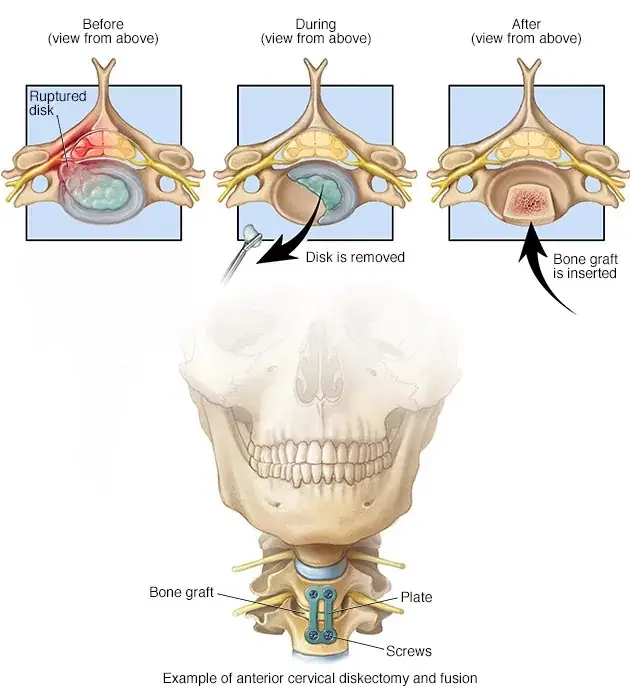
Anterior Cervical Discectomy and Fusion Surgery
Anterior cervical discectomy and fusion surgery (ACDF) is a procedure performed to reduce pressure on the spinal nerves and/or the spinal cord due to compression from a herniated disc or bone spur after all conservative treatments have been exhausted. This surgery is performed on the upper back/neck area and is two-fold, combining a discectomy and fusion surgery. It essentially consists of removing the damaged disc and then promoting bone growth and stability between the vertebrae.
ACDF surgery is a very common procedure and has a long and studied record of consistent positive outcomes. This is a highly successful surgery for pain relief with a very quick recovery. Disc replacement surgery is also available and appropriate in a select few cases.
Several factors will need to be considered prior to surgery, such as age, health status, lifestyle, anticipated levels of activity following surgery, as well as a number of appropriate diagnostic tests. Many patients present with complaints of neck and arm pain, caused by a herniated disc, cervical radiculopathy and spinal instability. Although many episodes of neck pain may be temporary, some patients experience chronic pain that ultimately requires surgery. There are three surgical options including cervical spinal fusion, cervical decompression surgery and disc replacement surgery.
You should be able to get up and walk around on the first day after the surgery. Dr Friesem will provide you with specific instructions including exercises to support your recovery.
?Why do you need Cervical (neck) Surgery
If you have been diagnosed with a herniated disc or bone spurs (which can form when the joints of the spine calcify) this can result in:
- Pain in the neck and/or arms
- Decreased balance
- You have been diagnosed with cervical degeneration requiring surgery
- Lack of coordination
- Numbness or weakness in the arms, forearms or fingers
- You have been diagnosed with compression of either the nerve roots in the cervical spine or compression of the spinal cord itself
- Painkillers, rest, exercises and injections have not alleviated your pain
- There is a likelihood of serious complications involving the nerves if left untreated
- The pain in your neck, upper back and arms is having a profound effect on your quality of life
?How is a Cervical Fusion Surgery performed
When cervical spinal fusion surgery is performed, the affected bones in the neck are ‘fused’ together using bone graft material with a plate and/or cage, so that they can heal together and form one healthy bone. Bone grafts can be taken from the patient’s pelvic bone, or cadaver and synthetic bone are also options.
Depending on the type of herniated disc and spinal surgery, Dr Friesem will use either an anterior approach (the spine is operated on from the front) or a posterior approach (the spine is operated on form the back).
Surgical Cervical Spinal Fusion Techniques
The method of your cervical fusion surgery will be discussed at length with you prior to the procedure and the decision of which method to use will be based on your unique condition, needs and surgical requirements.
- The bone graft is used to make a bridge between adjacent vertebrae and stimulates the growth of the vertebrae together
- Man-made fusion materials may also be used as opposed to taking bone from the patient’s own body or from a cadaver
- Metal implants can be used to hold the vertebrae together until new bone growth occurs (cages, plates, screws and/or rods)
- An entire vertebra can be removed and replaced with a cage-type device and the spine can then be fused
- A diseased or herniated spinal disc can be removed and replaced with an artificial device (disc replacement surgery)
Cervical Disc Replacement Surgery
Cervical disc replacement surgery involves removing a diseased cervical disc and replacing it with an artificial disc. Before this procedure was available, the affected disc was removed and the vertebrae above and below were fused together to prevent motion.
Disc replacement surgery has the advantage of allowing more movement and creating less stress on your remaining vertebrae than the more traditional cervical disc surgery.
?Why is Cervical Disc Replacement Surgery Needed
Loss of space between your cervical vertebrae from cervical disc degeneration, or wear and tear, is very common. Cervical discs begin to collapse and bulge with age. This happens to most people by age 60, but it is generally unknown why some people experience more pain and symptoms than others.
Symptoms may include
- Neck pain
- Neck stiffness
- Headache
- Pain that travels down into your shoulders or into your arms
- Weakness of your shoulders, arms, hands or legs
- Numbness or ‘pins and needles’ feeling in your arms
Surgical Procedure for Cervical Disc Replacement
The standard surgical procedure for a cervical disc replacement requires an anterior approach (from the front of the neck). A typical single-level surgery will take little more than an hour to perform.
- The surgery is performed using a general anesthetic and a 2.5-5 cm incision is made in the front of the neck.
- The affected disc is removed, as are any disc fragments or bone spurs that are pressing on a nerve root or the spinal cord.
- The disc space is restored to its normal disc height to help relieve pressure on the surrounding nerves.
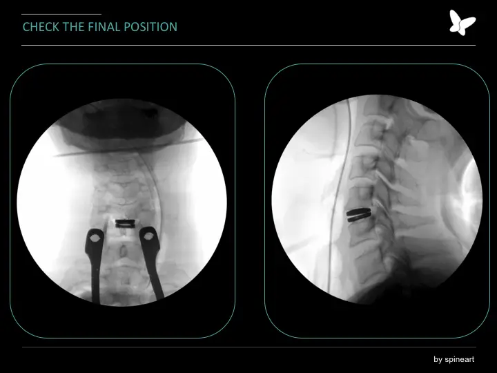
- Using live X-ray imaging for visual guidance, the artificial disc device is placed in the prepared disc space. More than one artificial disc size may be tried before the surgeon decides on the best fit.
- After the artificial disc is placed and attached to the 2 adjacent vertebrae (above and below), the incision is sewn up.
Lumbar Microdiscectomy
Microdiscectomy surgery may be performed after conservative treatments have not alleviated the patient’s painful symptoms. Microdiscectomy surgery involves removing a protruding or herniated disc and decompressing the lumbar nerves to relieve the pressure on the nerve roots. Microdiscectomy spine surgery decreases neural impingement and creates space, allowing the nerve to recover.
This fast, safe and effective spine surgery involves the use of microsurgical techniques (minimally invasive surgical techniques) to gain access to the lumbar spine – the surgical incision is typically about 2.5 cms long. Minimally invasive surgical technique refers to minimal damage to normal tissue. Only a small portion of the herniated disc that compresses the spinal needs to be removed. Microdiscectomy refers to the use of magnification for discectomy surgery. This allows minimal damage to soft tissues and muscles. Following microdiscectomy surgery, patients will be discharged home either on the same day or the following day (this may vary depending on each specific case).
A microdiscectomy is performed to alleviate the following painful conditions
- Sciatica – pain radiating down the back or side of the leg
- Herniated disc pain
- Weakness and/or numbness in the legs and/or feet
- Severe weakness due to chronic pain
- Debilitating leg pain
- Severe Femoratica (pain down the front of the thigh)
- Bowel or bladder incontinence
The results of microdiscectomy surgery have a relatively high success rate and compared to other spinal surgeries the postoperative healing time is much faster. Many patients will be back at work within a few days to a week. Moreover, this spinal surgery has a low complication rate.
Benefits of microdiscectomy surgery include:
- Quick procedure
- Minimal muscle and soft tissue disruption
- Short recovery time
- Minimal postoperative pain and discomfort
- Fewer risks of complications
Lumbar Spinal Fusion
Spinal fusion is a surgical procedure that is only recommended after conservative treatment methods have failed. There are a significant amount of factors that will be considered prior to Dr. Friesem’s recommendation to proceed with spinal fusion surgery.
Please note: Not everybody with lower back pain will require lumbar fusion surgery. However, if your back pain condition is one that requires surgery, this procedure has a long history as a safe and effective option.
Spinal fusion surgery involves using specialized instrumentation to perform a bone graft and permanently connecting two or more vertebrae together. This type of surgery is performed to provide stability and strength to the vertebrae and to eliminate chronic pain. Spinal fusion is actually one of the most common types of back surgery.
:Reasons Lumbar Spinal Fusion Surgery is performed
- Persistent back pain that has not responded to conservative methods – usually attributed to degenerative disc disease or spinal instability
- A definitive diagnosis of spinal stenosis (where there is an associated deformity)
- A definitive diagnosis of sponylolisthesis
- Prior failed laminectomy surgery
- Abnormal curvatures of the spine, such as scoliosis or kyphosis
- To correct a narrowing of the spinal canal
- Trauma to the spinal vertebrae – one or more fractured vertebrae
- To correct a spinal fracture
- Protrusion of the disc between the vertebrae (slipped disc, herniated nucleus pulposus)
- Weak or unstable spine caused by infection or tumour
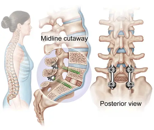
There are various spinal fusion techniques including a posterior approach (an incision in the lower back) or an anterior approach (an incision in the abdomen). Sometimes one approach is preferable to another depending on the MRI findings and the age, sex and health of the patient. The types of approach will be thoroughly discussed with the patient prior to surgery.
?What exactly happens during Lumbar Spinal Fusion Surgery
During spinal fusion (which occurs while the patient is under general anesthetic), Dr Friesem will make an incision either directly over the spine or in the abdomen (as previously mentioned, the exact method will vary from case to case and will be thoroughly discussed with you prior to surgery). A bone graft taken from your own body, from a bone bank or synthetic bone is placed between the vertebrae. Metal screws, rods or cages are inserted to stabilize the vertebrae while the bone graft heals.
:The goals of Lumbar Spinal Fusion Surgery include
- Eliminating neurological problems
- Relieving chronic pain (lower back and leg)
- Reducing further weakness
- Restoring healthy nerve function
- Stopping or preventing abnormal motion in the spine
- Realigning the spine to improve posture
Kyphoplasty
Kyphoplasty is a way of treating vertebral body compression fractures, which are small breaks in the thick mass of bone that makes up the front part of your spinal column (the vertebral body). Vertebral body fractures lead to the collapse or compression of a vertebra, causing your spine to shorten and curve forward. This can result in pain and a kyphotic (hunched-over) deformity.
Thinning of bones, or osteoporosis, is the main cause of vertebral compression fractures. Pathologic fractures related to spinal tumors may also be a cause.
Kyphoplasty is generally reserved for people with painful progressive (increasing) back pain caused by osteoporotic or pathologic vertebral compression fractures. Candidates for these procedures often have a reduced ability to move and function because of the fractures.
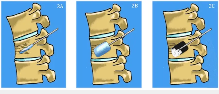
To be a candidate for a kyphoplasty your pain must be related to the vertebral fracture, and must not be due to other problems, such as disk herniation, arthritis, or stenosis (narrowing). Imaging tests — such as spinal x-rays, bone scans and computed tomography (CT) or magnetic resonance imaging (MRI) scans — might be ordered to confirm the presence of a vertebral fracture. If you have osteoporosis, Dr. Friesem may ask for a dual energy x-ray absorptiometry (DXA) scan.
The procedure is typically performed as an outpatient procedure. You’ll be sent home the same day.
After the bone cement is inserted into the vertebra, 75% of patients are active again. You might be symptom free and you won’t have to do physical therapy or rehabilitation.